Kidney Autoimmune Nephropathy Nephritis – IgA / Berger’s disease- Lupus – Immune Complex Recovery Protocols >>
Kidney Nephropathy IgA – Berger’s Disease Recovery Protocols >> LABs ….
—– 24 hour urine Creatinine Clearance
— Blood Immunoglobulin electrophoresis
— PAI1 – CRP high sensitivity
—- ANA and stippling pattern – Single and Double DNA antibody patterns etc. inflammatory markers
Plasmapheresis regularly with Immunologist at hospital if confirmed diagnosis
CORE PROTOCOLS >>
AllerGONE two capsules three times per day
AllerBlock two capsules three times per day
Cell Defense two caplets three times per day
Nutrimmune two capsules twice per day
Full Vitamin K2 two capsules twice per day
Power C PLUS four capsules three to four times per day
AllicinMed one capsule three times per day
ADVANCED PROTOCOLS >>
InFlamX three to four capsules three times per day
NutriTRALA one tablet three times per day
Cell Detox Glutathione two caplets twice to three times per day
SuperNOX with Beets three teaspoons in filtered water twice per day
Cell Defense Plus one capsule three times per day
NutriDine 5 drops in filtered water three times per day
COMPLETE PROTOCOLS >>
NutriDefense two capsules three times per day
OmegaSupreme PRO two softgels three times per day
MalignaBlock one capsule twice per day
Technologies >>
Lumen Photon Setting NICO 30 to 60 minutes per day after 15 to 30 minutes Setting 7 two to three times per day


Glomerulonephritis
Acute Post-Streptococcal Glomerulonephritis
Nephrotic Syndrome
Acute Post-Streptococcal Glomerulonephritis
-Post-infection with Group A Streptococcus
-Route respiratory; Skin injury
-Most common in school-age children
-Rare in infant
Pathophysiology
-Development of IgG 2-3 weeks after infection
-Antigen-antibody complex formation
-Deposited on glomerular capillary wall
-Activation of complement system
-Proliferation of mesangial and epithelial cells
-Infiltration of polymorphonuclear leukocytes in mesangium
-Renal biopsy–>Immunofluorescence–>IgG and C3 deposits
Clinical Features
-Ranging from asymptomatic –> heart failure; hypertensive encephalopathy
-Presentations: Malaise, anorexia, oligouria, smoky/brownish urine, facial puffiness, ankle oedema.
-May: Hypertension: Headache, vomiting, visual disturbance. Cardiac failure: cough, dyspnoea, chest pain, tachycardia, frothy bloo-stained sputum. Loin pain.
-Elevation of serum urea and creatinine
Management
+Bed Rest
+Antibiotics – 10 days penicillin
+Diet – salt restriction, protein restriction (1g/kg/day)
+Severe hypertension/encephalopathy
-diastolic P > 100mmHg
-antihypertensives: nefedipine, hydralazine, diazoxide
+Acute renal failure
-usually resolves within 2-3 weeks
-intravenous frusemide (severe oliguria, electrolyte and acid-base imbalance)
+Acute cardiac failure (left ventricule) – rare
Nephrotic Syndrome
-Clinical syndrome with gross proteinuria –> hypoalbuminaemia, oedema, ascites. Hyperlipidaemia usually present.
-Primary and Secondary causes:
Primary:
*Minimal change lesion
*Focal Segmental Glomerulosclerosis
*Mesangial Proliferative Glomerulonephritis
*Membrano-Proliferative Glomerulonephritis
*Membraneous Glomerulonephritis
*Congenital Nephrotic Syndrome
Secondary:
*Infection: Syphilis, Toxoplasmosis, Hep B, Post-streptococcal Glomerulonephritis, bacterila endocarditis, Quartan malaria.
*Drugs: Penicillamine, troxidone, heavy metals.
*Systemic diseases: Henoch-Schonlein purpura, SLE, long-standing DM.
*Malignancies: Hodgkin’s disease, leukaemia, lymphosarcoma.
*Others: CCF, Renal vein obstruction.
Minimal Change Lesion
-Commonest
-Male preponderance
-Mainly < 6years
-80% of nephrotic syndrome < 15 years of age
Pathophysiology
-T-cell abnormalities
-Production of lymphokine
-Increases permeability of basement membrane
-Normal Light Microscopy & Immunofluorescence
-Swelling + Fusion of foot processes of epithelial cells on Electron microscopy
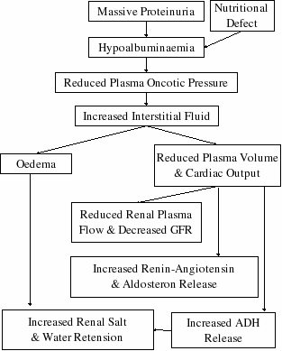
Pathogenesis of oedema in Nephrotic Syndrome

Clinical Features
-Gradual onset
-Facial puffiness, ankle oedema, abdominal distension –> whole body swelling.
-Oedema in morning –> subsides in evening
-Urine clear & frothy
-Albuminuria +++ to ++++
-Usually BP normal, no hematuria
-Serum Albumin < 25g/L
-Elevated serum Cholesterol
-Nomal serum urea & creatinine
-GFR normal
-Minor: Hypertension, microscopic hematuria, reduced GFR (hypovolaemia)
Management
+Bed rest – severe gross oedema
+2mg/kg/day prednisolone –> remission + 2-4 weeeks continuation – 2/3 divided doses
+Remission is ‘complete loss of proteinuria, serum Albumin > 24g/L
+Then 1.5mg/kg/day single morning dose / 2mg/kg/day alternate day for 4 weeks
+Cyclophosphamide 3mg/kg/day single morning dose (if dev side effect to steroid)
Complications
1. Infections
-peritonitis, pneumonia, cellulitis & septicaemia
-give broad spectrum antibiotic
2. Severe hypovolaemia
-suspected if grossly oedematous, abdominal pain, vomiting, rapid pulses, cold extremities.
-serum Albumin < 12g/L
3. Respiratory embarrassment
-due to pleural effusion, severe ascites
4. Hypercoagulable state
-due to haemoconcentration , loss of blood factors –> give aspirin
Refer Table for differences between Minimal Change NS & Significant Glomerular Lesions NS
Focal Segmental Glomerulosclerosis (FSGS)
-5-10% of NS
-Same clinical Presentation as Minimal Change NS
-10-15% response to steroid initially, later become steroid resistant
-May present with hypertension, haematuria, casts.
-Biopsy: Focal areas of segmental sclerosis.
-Immunofluorescence: Hyalinosis with IgM & IgG.
-Later develop chronic renal failure –> end stage renal disease in 10 years.
Membrano-proliferative Glomerulonephritis
-8% of NS
-Usually gross haematuria.
-May present as Acute nephritis.
Mesangial Proliferative Glomerulonephritis
-Usually resistant to steroid. Can have partial response.
-Frequent episodes of heavy proteinuria with severe oedema.
Membranous Glomerulonephritis
-Rare.
-Can present as asymtomatic proteinuria.
-Some response to steroid, but rarely remit completely.
-Associated with Syphylis, Toxoplasmosis, Hepatitis B, Malignancies.
Congenital NS
-1st few month of life.
-Causes:
-Intrauterine infection: Syphylis, Toxoplasmosis.
-Renal vein thrombosis.
-Finnish type Congenital NS:
-High level of alpha-fetoprotein in amniotic fluid.
-100% mortality without renal transplant.

